If you’re a nurse practitioner new grad, documenting a patient encounter can be a bit of a struggle. You need to present your patient in a manner such that other providers can glance at the chart and pick up where you left off. You must also provide sufficient documentation so that you can justify your decision making should questions about the care you provided arise. Completing charts in a time efficient manner is no easy task, particularly early in your nurse practitioner career.
That is why charting is a key component in our transition to practice curriculum. To give you a feel for the skills you will develop in the ThriveAP virtual NP program we’ve been doing a system-by-system series on documentation. These posts will help guide your charting giving you the words you may need to accurately represent a physical exam. Today, let’s check out basic documentation of the HEENT (Head, Eyes, Ears, Nose, Throat) exam.
What You’re Looking For
Whether you’re doing a well child check up, or assessing a patient for an acute illness, as a nurse practitioner you’ll likely do your fair share of HEENT exams and must be familiar with the exam’s basic components.
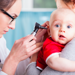
- Inspection – Assess the head and face for external abnormalities such as erythema in the presence of infection, or bruising and deformity in the event of trauma. In the HEENT exam, inspection also includes visualization of the ear canals and tympanic membrane with an otoscope as well as the pharynx.
- Palpation – Palpate for areas of tenderness or other abnormalities such as masses or lymphadenopathy.
While assessing additional items like sensation of the face are important, we have covered these in the documentation review of the neurological system.
Buzzwords to Know
There are a few terms you may encounter in HEENT documentation that can be helpful to include in your own charts. Familiarize yourself with these terms as you perfect your exam and documentation technique.
- EOM (Extraocular Movements) – Movement of the eye in all directions, up, down, right and left. These movements are controlled by a set of small muscles in the face. Entrapment of these muscles can occur, particularly with trauma, causing problems with ocular movement.
- PERRLA – Shorthand for Pupils Equal, Round, React to Light, Accommodation. Helpful for documenting an eye assessment.
- Nystagmus – Visual condition in which the eyes make repetitive and uncontrolled movements.
Sample Normal Exam Documentation
Documenting a normal exam of the head, eyes, ears, nose and throat should look something along the lines of the following:
Head – The head is normocephalic and atraumatic without tenderness, visible or palpable masses, depressions, or scarring. Hair is of normal texture and evenly distributed.
Eyes – Visual acuity is intact. Conjunctivae are clear without exudates or hemorrhage. Sclera is non-icteric. EOM are intact, PERRLA. Fundi appear normal including optic discs and vessels. No signs of nystagmus.
Ears – The pinna, tragus, and ear canal are non-tender and without swelling. The ear canal is clear without discharge. The tympanic membrane is normal in appearance with a good cone of light. Hearing is intact with good acuity to whispered voice.
Nose – Nasal mucosa is pink and moist. The nasal septum is midline. Nares are patent bilaterally.
Throat/Mouth – Oral mucosa is pink and moist with good dentition. Tongue normal in appearance without lesions and with good symmetrical movement. No buccal nodules or lesions are noted. The pharynx is normal in appearance without tonsillar swelling or exudates. No adenopathy is noted.
Sample Abnormal Exam Documentation
When documenting an exam abnormality, be as specific as possible about where the abnormality lies and what the abnormality looks like. You will not document all of these abnormalities in a single exam, but the following are some HEENT abnormalities you may need to include in your documentation.
Head
- Microcephalic or macrocephalic
- Temopral artery tenderness
- Swelling, lesions, or tenderness to scalp
- Abnormalities to external appearance of face and/or asymmetry
Eyes
- Ptosis (drooping of upper eyelid)
- Exopthalmos (protruding eyes) or enopthalmus (sunken eyes)
- Conjunctival erythema or hemorrhage
- Scleral icterus
- Drainage
- Pupils – constricted or dilated
- Nystagmus
- Foreign body
- Eyelids – swelling, erythema, hordeolum (stye)
Ears
- External tenderness
- Swelling, lesions, drainage
- Tympanic membrane – erythema, perforation, bulging, effusion
- Ear canal – swelling, exudates, foreign body
Nose
- Lesions, swelling, or asymmetry
- Signs of trauma – bruising or swelling
- Discharge
- Flaring (in respiratory distress)
- Septal deviation
- Foreign body
Throat/Mouth
- Cracking or signs of dehydration to lips
- Missing, loose, decayed, or abnormally positioned teeth
- Intraoral lesions or sores
- Swelling and/or exudate to tonsils
Remember, these systems don’t exist in isolation. For example, you may need to incorporate a respiratory exam, or document additional findings such as lymphadenopathy relating to your exam. The depth with which you examen and chart on the head, eyes, ears, nose, and throat depends on the patient’s presentation and history.
Learn more about the ThriveAP transition to practice education and how you can leverage the program to accelerate your NP career.


